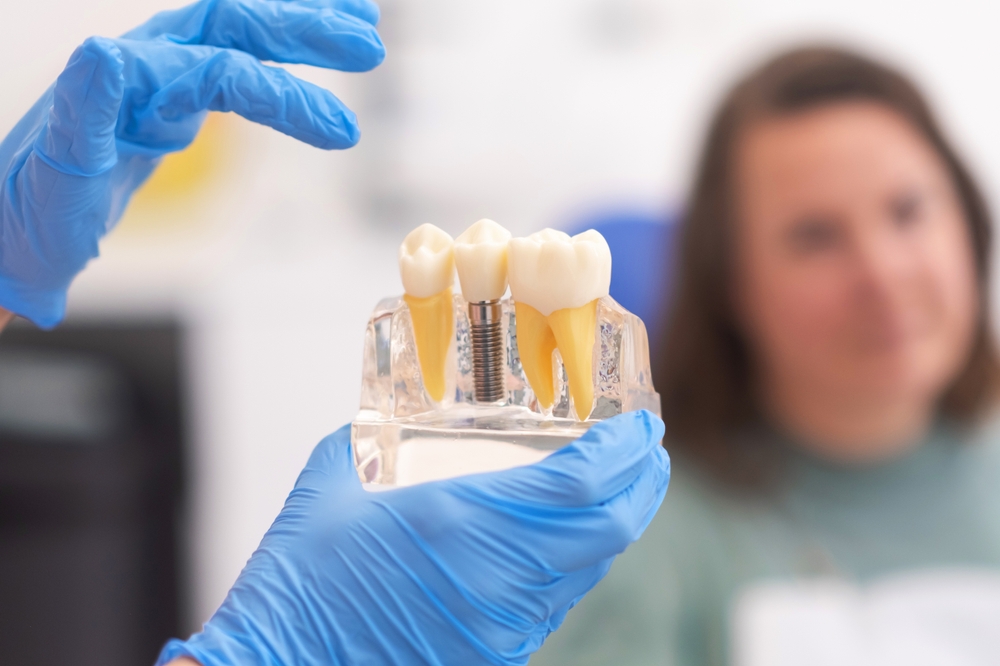
The history of dental implants to replace missing teeth begins in ancient Egyptian time approximately 2500 BC. They used ligature wire made of gold and used artificial teeth from animal bones to replace missing teeth. The Etruscans in 500 BC made teeth from oxen bones. The Phoenicians used gold wire and teeth were carved out of ivory in 300 AD. The Mayan population excelled in replacement of missing teeth. They used shells as implants. Around 800 AD the Honduran culture placed stones in the mandible for missing teeth.
In more modern history, in Europe from 1500 to 1800, teeth were collected from cadavers for allotransplantation. In the 1930’s, latticed cylinders were placed in the mandible. This technique was continued, and a threaded cap was ultimately developed and placed over the implant.
The Father of Modern Implantology, Formiggini, developed the stainless steel design of the implant that allowed bone to grow into the metal. Finally, Dr. Andres from Spain modified Formiggini’s spiral design to include a solid shaft in the construction.
Many design changes have been made over the modern era. One of the main reasons for the modifications of the implants was made to decrease the healing time . The surface of a dental implant is the only part that is in contact with the bio-environment and the uniqueness of the surface directs the response and affects mechanical strength of the implant/tissue surface. The surface treatment layer on the implant is required to increase the functional surface area of the implant /bone interface so stress can be effectively transferred.
These surface treatments were found to control the growth and metabolic action of osteoblasts. Also, surface roughness has been shown to influence cytokine and growth factor production by osteoblasts. Increased roughness allowed transforming growth factor beta production which directly increased osteoblastic propagation. This suggests that the structure of the implant influenced the interaction between the metal and the living tissue.
Complications of Dental Implants
Failure of osseointegration is one of the main complications. Osseointegration is when the bone grows around the implant. If this process fails or is only partially grown, there will be failure of the implants as it will not be secure enough to handle the stress. The implant will either fall out or will need to be removed.
Improper implant placement is another complication. If there is a problem with the location or angle of the implant it will cause problems. Fusion may not occur properly, and painful chewing typically is reported.
Risks of Dental Implant Surgery
Swelling and bruising is common. This is self-limited and resolves with time.
Sinus damage can occur as the upper teeth can potentially penetrate into the sinus cavity when being replaced. This can lead to pain and infection.
Infection is a large risk. Some newer implants are coated with antibiotics to help minimize this risk.
Nerve damage can happen as there are many adjacent nerves.
Other teeth can be affected around the implant site and the trauma from the surgery can make them worse if they have decay or root damage.
Jaw preparation is a critical step in implant surgery. The jaw must be strong enough to hold the implant. Bone density is important and stimulation of continued bone growth around the implant is mandatory.
During surgery, the gum is incised, and the bony jaw is exposed. The implant is then inserted into the jaw. In some cases, the abutment which sits above the gum line is attached. The artificial tooth attaches to this post.
What does Hyaluronic Acid have to do with this procedure?
Hyaluronic acid is a linear glycosaminoglycan found in the extracellular matrix of epithelial and connective tissue of vertebrates. As a major component of the extracellular matrix, hyaluronic acid has a key role in tissue regeneration, inflammation response and angiogenesis.
Granulation tissue is the perfused, fibrous connective tissue that replaces a fibrin clot in healing wounds. It typically grows from the base of an injury and expands as the wound heals. Hyaluronic acid is abundant in the granulation tissue matrix. This HA-fibrin network has a variety of functions in tissue repair. These functions include cell migration to the site, cell proliferation and organization of the tissue matrix. Inflammation is critical for the healing process and the pro-inflammatory role of HA contributes to the overall healing cascade.
Platelet Rich Fibrin
Platelet Rich Fibrin is called the “second generation of platelet products”. In its simplest form, platelet rich fibrin is a fibrin membrane with trapped platelets and leukocytes. Included in the PRF are cytokines, growth factors, bioactive proteins and adhesive proteins such as fibronectin and fibrinogen. Leukocytes are present not only for their anti-infective actions, but they also secrete a large quantity of growth factors. The polymerization of fibrin produces a 3-dimensional cross linked fibrin matrix. This serves as the binding site for other platelets and growth factors. By increasing growth factors at a specific tissue location, PRF promotes tissue regeneration by mimicking the wound repair process over a prolonged period of time. Platelet Rich Fibrin is anti-inflammatory and has cytokines and growth factors that stimulate the repair mechanism of tissue such as the stimulatory enhancement of osteoblasts and fibroblasts in the dental environment.
Studies
Choukroum was the first to use platelet rich fibrin in dentistry. His published paper in 2006, was on histologic generation of PRF effects of bone allograft maturation in sinus lift procedures. He showed that freeze dried bone allograft combined with PRF increased bone formation in this procedure. Many future surgical uses for PRF in dentistry followed especially to facilitate periodontal intrabony defects.
Clin Oral Investig 2017 Nov;21(8):2619-2627 PMID 28154995
Objective: Platelet Rich Plasma has been utilized in regenerative dentistry as a supra-physiological concentrate of autologous growth factors capable of stimulating tissue regeneration. In this study, a liquid formulation of platelet rich fibrin termed injectable platelet rich fibrin without anti-coagulants was investigated.
Conclusion: Injectable Platelet Rich Fibrin demonstrated the ability to release higher concentrations of various growth factors and induced fibroblast migration and expression of PDGF, TGF-B, and collagen.
Odontology 2024 May 21 PMID 38771493
Objective: One of the most promising approaches to correct periodontal bone defects and achieve periodontal regeneration is platelet rich fibrin. This review and meta-analysis aimed to evaluate the regeneration of periodontal bony defects using PRF compared to other regenerative treatments.
Conclusion: The use of platelet rich fibrin significantly improved the regeneration of periodontal bone defects compared to other treatments.
J Appl Oral Sci 2024 May 10:32;e20230294 PMID 38747782
Objective: This study aims to develop a compound biomaterial to achieve effective soft tissue regeneration
Conclusion: The HA-PRF group exhibited greater potential to promote soft tissue regeneration by inducing cell proliferation, collagen synthesis and migration of growth factors than hyaluronic acid or platelet rich fibrin group. HA-PRF can serve as a great candidate for use alone or in combination with autografts in periodontal or peri-implant soft tissue regeneration.
Juventix Regenerative Medical is an industry leader in the regenerative medical field. Our Platelet Rich plasma Kits are FDA cleared and designed for safety, sterility and effectiveness. Our kits are scientifically manufactured to provide a platelet concentrate, devoid of red blood cells with a minimum number of leukocytes that are critical to the regenerative process.
Juventix Regenerative Medical offers a LED Activator to activate the platelets and begin the regenerative process. The activation is a critical step in the release of growth factors, cytokines and bioactive proteins from the alpha granules on the platelets and is accomplished with LED light. This negates the use of chemicals such as Calcium Chloride, Thrombin or Collagen. This mode of activation by LED light provides sustained growth factor release versus older methods of activation while adhering to the minimal manipulation guidelines of the FDA.
Juventix Regenerative Medical supplies a Bio-Incubator to transform the Platelet Rich Plasma into an Injectable Platelet Rich Fibrin. The Platelet Rich Fibrin, commonly called the “second generation of platelet products” has different growth factors and cytokines than the original PRP. These cytokines provide an anti-inflammatory microenvironment and can be used confidently in inflammatory conditions.
Juventix Regenerative Medical has a vast array of products, services and devices for the regenerative and dental professional. Always striving to providing the cutting edge, FDA cleared products at a cost-effective price.
RESTORE, REVIVE, REGENERATE- JUVENTIX REGENERATIVE MEDICAL
Regenerative Regards,
Dr. Robert McGrath


